- Usage
- Usage Cases
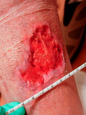
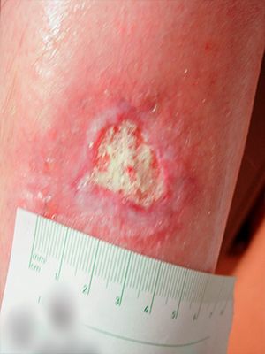
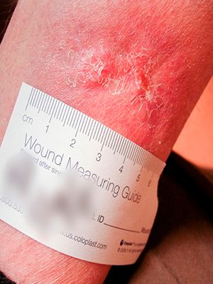
- Venous Leg Ulcer
Case №31
Venous Leg Ulcer
Age of Wound: 3 months
USA
-
Gender:
Man
-
Age:
62 years
-
Diagnosis:
Venous Leg Ulcer
Types of trophic ulcers

The types of trophic ulcers are most often isolated for the reasons of their occurrence, that is, for the initial diseases: venous, atherosclerotic, diabetic, neurotrophic, hypertensive and infectious. Let's try to briefly describe each of the types.
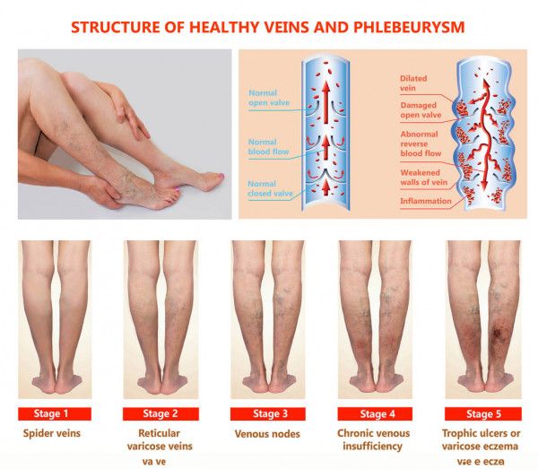
Venous ulcers are a complication of varicose veins in violation of the blood supply to the lower extremities. Most often, lesions occur on the lower parts of the inner surface of the legs. The severity and cramps progress to the stage of itching and the formation of a "mesh" of veins. Further, the skin becomes denser, smoothed, and shines. The next unpleasant moment is the formation of whitish clamps. Start treatment urgently! After all, literally in a few days an ulcer forms on the skin, which is prone to progression and can lead to sad consequences.
Atherosclerotic ulcers are a consequence of ischemia of the soft tissues of the lower leg. After all, obliterating atherosclerosis affects the arteries. Tight shoes or hypothermia can cause the formation of a trophic ulcer on the foot. This is a semicircular purulent fossa. The patient has cold feet, pains at night, it is difficult to climb stairs - more often these are elderly people.
Diabetic ulcers are a complication of diabetes. They begin with the death of nerve endings and a decrease in the sensitivity of the skin of the legs. After some time, pains are added, especially at night. An ulcer is often located on the big toe due to injuries to the calluses of the foot. Because of the high likelihood of infection, such an ulcer can lead to amputation.
Neurotrophic ulcers are ulcers due to head or spinal trauma that appear on the sole or lateral part of the heel. A deep, but small crater, reaching the muscles and even bones, in which pus accumulates.
Hypertensive ulcers are a consequence of prolonged spasm of the walls of small vessels against the background of a very long period of high blood pressure. This is an extremely rare species that can nevertheless occur in women after 40 years. The main differences between this type of ulcer are symmetrical arrangement on the outer surface of the legs, slow development and severe pain.
Infectious ulcers are the consequences of reduced immunity and lack of hygiene. With this type of trophic lesions, single oval ulcers and small depths or groups of them appear over the entire surface of the legs.
Cooperate With Us
or via contact phones or email
-
Conveniently
By contacting only one supplier, our customers receive a wide range of products in the shortest possible time.
-
Simple
Our customers are only required to form an order for the supply of products, we will take care of all further tasks
-
Profitable
We save our customers money on import
and delivery taxes


