- Usage
- Usage Cases
- Polytrauma
Case №52
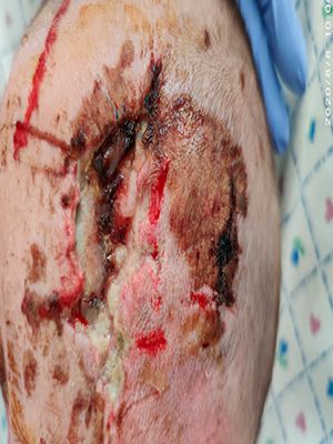
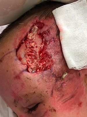
Polytrauma, TMT, open fracture of the frontal bone on the left, with a transition to the superior wall of the orbit, without damage to the dura mater and structures of the left eye, extensive laceration of the left fronto-parietal region, chest contusion
Age period of the presence of a wound: 20 days
Attending physician: Professor Marina Georgievna Melnichenko, DebigaVyacheslav Valerievich
Regional Children's Clinical Hospital, Dept. neurosurgery, Odessa
-
Patient:
Boy
-
Age:
1 year 7 months
-
Diagnosis:
Polytrauma
Traumatic brain injury
Traumatic brain injury - headache, tinnitus, dizziness, nausea, vomiting, loss of consciousness and memory is possible. In such cases, emergency specialized medical care is required.
Classification
According to the severity of the lesion, there are mild, moderate and severe TBI. The Glasgow Coma Scale is used to determine the severity. In this case, the patient receives from 3 to 15 points depending on the level of impairment of consciousness, which is assessed by opening the eyes, speech and motor responses to stimuli. Mild TBI is estimated at 13-15 points, moderate - at 9-12, severe - at 3-8.
They also distinguish between isolated, combined (injury is accompanied by damage to other organs) and combined (various traumatic factors act on the body) TBI.
TBI is divided into closed and open. With an open traumatic brain injury, the skin is damaged, the aponeurosis and the bottom of the wound is bone or deeper tissues. Moreover, if the dura mater is damaged, then an open wound is considered penetrating. A special case of penetrating trauma is the leakage of cerebrospinal fluid from the nose or ear as a result of a fracture of the bones of the base of the skull. In a closed traumatic brain injury, the aponeurosis is not damaged, although the skin may be damaged.
Clinical forms of TBI:
Fracture of the bones of the skull - Fractures are more often bone-linear.
- Concussion is a trauma-induced impairment of neurological function. All symptoms that occur after a concussion usually disappear over time (within a few days - 7-10 days).
Persistent persistence of symptoms is a sign of more serious brain damage. Concussion may or may not be accompanied by loss of consciousness. The main criteria for the severity of a concussion are the duration (from a few seconds to 5 (in some sources up to 20) minutes) and the subsequent depth of loss of consciousness and a state of amnesia.
Nonspecific symptoms - nausea, vomiting, pallor of the skin, cardiac disorders. Neurologic examination usually shows no abnormalities, but somatic symptoms (headache), physical signs (loss of consciousness, amnesia), behavioral changes, cognitive impairment, or sleep disturbance may occur. Some of these effects can last several months after the injury.
Brain contusion: mild, moderate and severe (clinically). Brain contusion manifests itself in a bruised wound of the brain tissue. A bruise, a shock-shock, is inflicted when the brain hits the skull wall at the point of direct impact of an external object on the head, receives one bruised wound and then the bruised wound is inflicted on the opposite side of the brain with a sharp slowdown in the movement of brain tissue. Clinical manifestations depend on the location of the injury, and include a change in mental state, increased drowsiness, confusion, agitation.Minor intraparenchymal hemorrhages and swelling of the surrounding tissue can often be identified with computed tomography.
Impact and counter-impact injury
Diffuse axonal injury - severe damage to the axonal white matter of the brain due to shear force caused by strong acceleration or deceleration of the brain
- Compression of the brain
Intracranial hemorrhage (hemorrhage in the cranial cavity: Subarachnoid hemorrhage, Subdural hematoma, Epidural hematoma, Intracerebral hemorrhage, Ventricular hemorrhage)
Combination
At the same time, various combinations of types of traumatic brain injury can be observed: contusion and compression by hematoma, contusion and subarachnoid hemorrhage, diffuse axonal damage and contusion, contusion of the brain with compression by hematoma and subarachnoid hemorrhage.
Symptoms
- damage to the cranial nerves, indicate compression and contusion of the brain.
- focal lesions of the brain indicate damage to a certain area of the brain, occur with contusion, compression of the brain.
- stem symptoms - are a sign of compression and bruising of the brain.
- Meningeal symptoms - their presence indicates the presence of a brain contusion, or subarachnoid hemorrhage. Several days after the injury may be a sign of meningitis that has developed.
Clinical picture
External hematomas, tissue ruptures
- pallor;
- vomiting, nausea;
- irritability;
- lethargy, drowsiness;
- Weakness;
- paresthesia;
- headache;
- impairment of consciousness - loss of consciousness, somnolence, stupor, coma, amnesia, confusion;
- neurological signs - convulsions, ataxia;
The main indicators of the state of the body may change - deep or arrhythmic breathing, hypertension, bradycardia - which may indicate increased intracranial pressure
Cooperate With Us
or via contact phones or email
-
Conveniently
By contacting only one supplier, our customers receive a wide range of products in the shortest possible time.
-
Simple
Our customers are only required to form an order for the supply of products, we will take care of all further tasks
-
Profitable
We save our customers money on import
and delivery taxes


